


Flow Cytometry and Cell Separation Unit
Flow cytometry, as a quantitative multiparametric analysis technique, is an interdisciplinary tool that makes it possible to quantify cell populations of interest rapidly, sensitively, objectively and simultaneously, based on the principles of fluorescence and light scattering.
This technology is based on passing cells or other suspended particles, aligned within a laminar flow, through a flow chamber illuminated by a coherent light beam at a specific wavelength. The optical properties measured for each cell are related to light scattering and to the fluorescence emitted by the cells when excited by the light beam. The use of fluorochromes with excitation wavelengths compatible with the cytometer's light sources and with different emission spectra makes it possible to study simultaneously different biological parameters and the functional and phenotypic characteristics of individual cells, even in heterogeneous samples.
The technological advances implemented in today's high-capacity flow cytometers make this analytical method highly applicable in basic research, clinical research and clinical diagnostics.
The objectives of the IDIPHIM Flow Cytometry and Cell Sorting Unit are:
- To provide technical support and advice to the centre's research groups and visiting researchers on experimental design, marker selection, sample acquisition, cell population analysis and discussion of the results obtained.
- Isolation of cell populations using cell-sorting technology.
- Development of new technologies and evaluation of new equipment.
- Organisation of training courses in this technology for users.
The Flow Cytometry and Cell Sorting Unit of the Puerta de Hierro Health Research Institute is equipped with state-of-the-art scientific equipment for research and is suitable for carrying out all its functions as a support unit for both internal and external researchers.
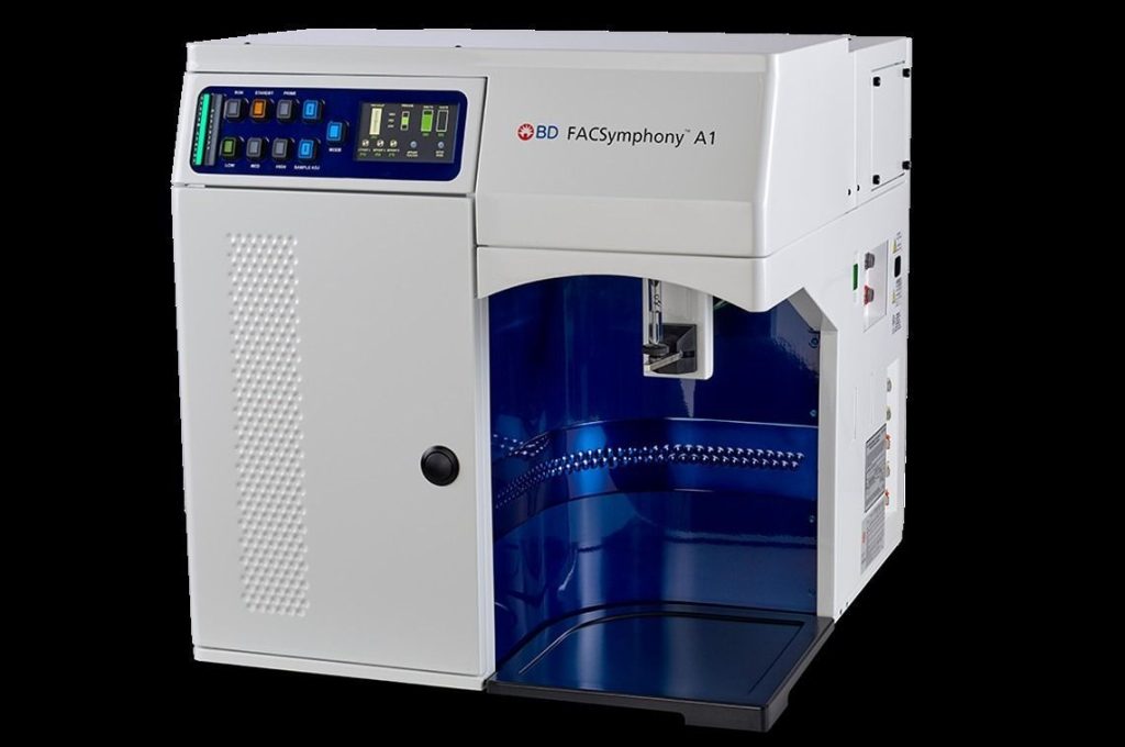
FACSYMPHONY A1 ANALYTICAL FLOW CYTOMETER
The Flow Cytometry and Cell Sorting Unit provides users with a HIGH-PERFORMANCE 4-LASER ANALYTICAL FLOW CYTOMETER (BD) capable of detecting 19 parameters simultaneously (16 fluorescence and 3 light-scatter parameters). It was funded under the State Subprogramme for Scientific and Technical Infrastructure and Equipment of the 2021–2023 Strategic Action in Health, charged to European funds from the Recovery, Transformation and Resilience Plan. (IFEQ22/00172).
The Flow Cytometry and Cell Sorting Unit provides users with a HIGH-PERFORMANCE 4-LASER ANALYTICAL FLOW CYTOMETER (BD) capable of detecting 19 parameters simultaneously (16 fluorescence and 3 light-scatter parameters). It was funded under the State Subprogramme for Scientific and Technical Infrastructure and Equipment of the 2021–2023 Strategic Action in Health, charged to European funds from the Recovery, Transformation and Resilience Plan. (IFEQ22/00172).
FACSARIA II CELL SORTER
High-speed digital FACSAria II cell sorter (BD) capable of sorting up to four phenotypically distinct cell populations simultaneously under both sterile and non-sterile conditions. It has two lasers at 488 nm and 633 nm and can detect 7 fluorescence parameters in addition to parameters relating to size (FSC) and cell complexity (SSC).
High-speed digital FACSAria II cell sorter (BD) capable of sorting up to four phenotypically distinct cell populations simultaneously under both sterile and non-sterile conditions. It has two lasers at 488 nm and 633 nm and can detect 7 fluorescence parameters in addition to parameters relating to size (FSC) and cell complexity (SSC).
AUTOMACS PRO MAGNETIC CELL SEPARATOR
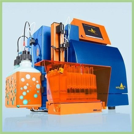
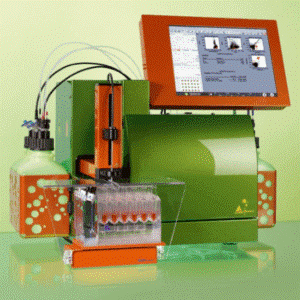
The AUTOMACS PRO system (Miltenyi Biotec) enables cell populations to be isolated from biological samples using immunomagnetic methods. The AUTOMACS PRO system can process up to 6 samples at a time using different selection procedures, with each sample programmed automatically in advance.
The AUTOMACS PRO system (Miltenyi Biotec) enables cell populations to be isolated from biological samples using immunomagnetic methods. The AUTOMACS PRO system can process up to 6 samples at a time using different selection procedures, with each sample programmed automatically in advance.
FC500 ANALYTICAL FLOW CYTOMETER
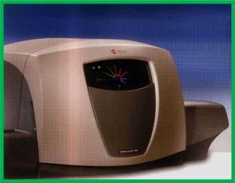
The Flow Cytometry and Cell Sorting Unit also provides users with an additional cytometer on loan from the Immunology Department. This is the FC500 flow cytometer (Beckman Coulter), with an optical configuration comprising a blue laser (488 nm) and 5 fluorescence detectors, enabling cell-biology studies to be carried out.
The Flow Cytometry and Cell Sorting Unit also provides users with an additional cytometer on loan from the Immunology Department. This is the FC500 flow cytometer (Beckman Coulter), with an optical configuration comprising a blue laser (488 nm) and 5 fluorescence detectors, enabling cell-biology studies to be carried out.
MACSQUANT 10 ANALYTICAL FLOW CYTOMETER
The Flow Cytometry Unit has a digital MACSQUANT analytical flow cytometer (MILTENYI BIOTEC) equipped with three air-cooled, fixed-alignment solid-state lasers: violet at 405 nm, blue at 488 nm and red at 635 nm. It enables the simultaneous detection of 10 parameters (8 optical channels for fluorescence measurements and 2 detectors for physical parameters relating to size (FSC) and cell complexity (SSC)). Fluorescence compensation is performed using an 8×8 inter- and intra-laser matrix system with automatic recalculation following voltage adjustments.
The Flow Cytometry Unit has a digital MACSQUANT analytical flow cytometer (MILTENYI BIOTEC) equipped with three air-cooled, fixed-alignment solid-state lasers: violet at 405 nm, blue at 488 nm and red at 635 nm. It enables the simultaneous detection of 10 parameters (8 optical channels for fluorescence measurements and 2 detectors for physical parameters relating to size (FSC) and cell complexity (SSC)). Fluorescence compensation is performed using an 8×8 inter- and intra-laser matrix system with automatic recalculation following voltage adjustments.
DATA ANALYSIS SOFTWARE
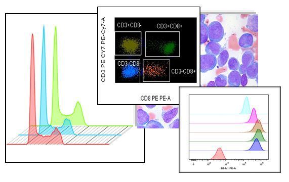
- FACSDiva software (BD): enables data acquisition on the FACSAria II cell sorter and subsequent analysis, with plots using linear, logarithmic and biexponential scales, as well as export of statistical data and templates.
- MACSQuantify software (Miltenyi Biotec): runs under Windows and provides all the functions required to display data, perform image and statistical analysis, analyse data during sample acquisition, group data and generate a single file. Custom analysis templates can also be created.
- FLOWJO software (BD): data-analysis software for data generated by any flow cytometer. It provides a range of analytical platforms that enable more specialised analyses to be performed.
- INFINICYT software (BD): includes tools for analysing, integrating and interpreting results, such as plots based on principal component analysis that provide information on the most significant markers for separating and identifying the cell populations under study; the ability to merge and integrate several files into a single file for global analysis; and the ability to create reference images.
TYPES OF TECHNIQUES USED
The following techniques are carried out at our facilities.
- Flow-cytometric quantification of immunophenotypic profiles: Preparation of biological samples for multicolour immunophenotyping of cell populations. This is performed using direct or indirect fluorescence techniques with monoclonal antibodies conjugated to fluorochromes for the characterisation of surface, intracellular or nuclear molecules that identify the phenotypic profile of populations of interest.
- Fluorescence-based cell sorting (FACS ARIA II) Multicolour phenotyping allows us to isolate pathological populations that are present at very low levels in a sample. This is based on the optical characteristics of the equipment and on its fluidics, which allow the physical separation, by electrostatic methods, of a specific population of interest from the overall cell suspension.
- Cell separation using the MACS immunomagnetic system: A column-based system in which the columns are placed inside a magnet. Cells are labelled with microbeads to identify epitopes of interest on the cell surface and are retained in the column. Populations that are not retained make up the negative fraction, containing cells without the ligand for the antibody used. This achieves high levels of purity. This automated system, based on coating the cells of interest with iron beads, enables their isolation while having minimal impact on the cells and maintaining high levels of viability.
- Tissue dissociation: using mechanical, enzymatic or automated dissociation systems, including the GentleMACS tissue dissociator (Miltenyi Biotec), to obtain cell suspensions that can subsequently be characterised with monoclonal antibodies for flow-cytometric quantification.
- Preparation of cell cultures for different procedures: functional studies by flow cytometry, metabolic studies (REDOX activity generation), and quantification of gene expression in living cells using fluorescent proteins.
- Cultured cells for the study of extracellular microparticles and their involvement in pathological processes as a source of potential therapeutic targets.
TYPES OF ASSAYS
The flow cytometry and cell sorting laboratory performs, among others, the following assays:
- Multicolour analysis of surface and intracellular cell antigens.
- Cell-viability assays: drug screening on cell lines. Identification of different stages of apoptotic cell death.
- Cell-cycle analysis. Proliferation assays (CFSE).
- Cell-signalling studies.
- Functional studies (drug resistance, kinetic assays, cell-permeability studies, detection of intracellular generation of reactive oxygen species (ROS) in cell lines and tissue samples).
- Reporter genes: analysis of fluorescent-protein expression efficiency.
- Cell sorting using FACS or immunomagnetic isolation.
- Detection of circulating tumour cells (CTCs) in peripheral blood and characterisation of antigen expression using multiparametric labelling.
- Studies of extracellular macroparticles and/or exosomes by quantification in the small-particle module and fluorescence side scatter (SPD).
Samples analysed
At the Flow Cytometry Unit we process and analyse biological samples from clinical trials and patients, PBMCs isolated for research studies, cell cultures, tissue dissociation, etc., for the characterisation and/or isolation of cell populations of interest. We have established control samples for quantifying parameters in each procedure and/or working protocol for each requesting user.
Requests must be submitted by completing the corresponding request form, which will be available on the Unit website:
- Data Analysis Service request form
- Flow Cytometry Service request form
- Cell Sorting Service request form
Sample-receipt conditions will be taken into account, including correct sample identification and presentation conditions, with particular consideration given to sample viability.
The information to be provided by the researcher in order to carry out an analytical flow-cytometry study is:
- Experimental design and number of samples.
- Protocol to be carried out.
- Cell type to be analysed.
- Combination of fluorochromes to be used in the experiment.
When requesting cell sorting of samples of interest, researchers must provide the following information:
- Experimental design.
- Protocol to be carried out.
- Cell type to be sorted.
- Number of samples to be sorted.
- Identification of the cell populations to be sorted: by fluorescence emission from the fluorochromes used (in the case of the FACSAria II sorter) or by immunomagnetic beads (when using the AUTOMACS PRO separator).
- Cell concentration of the sample to be sorted.
- Method for collecting the sorted cells (type of tube).
- Whether sterile conditions are required during sample processing.
The necessary cleanliness levels are maintained on all equipment and in the laboratory for assays requiring sterile conditions.
Data analyses are performed comparatively between control samples and samples of interest using standardised analysis software that is widely accepted internationally.
FLOW CYTOMETRY AND CELL SORTING UNIT FEES

STAFF AND LOCATION
- Scientific Head of the Unit: Dr Aránzazu García Grande
Tel.: 91 191 71 04 // 91 191 67 56
Email: citometria@idiphim.org - Laboratory technical staff: Marta Garcia Gil
- Location: 3rd floor of the Research Area, Research Institute, Hospital Puerta de Hierro Majadahonda
SERVICE BOOKING FOR PUERTA DE HIERRO RESEARCHERS:
To use the service, you must make an advance booking via the Unit website (an internal user password is required).
SERVICE BOOKING FOR EXTERNAL USERS:
You may request the service by contacting the Head of the Unit by email in advance.