

Advanced Optical and Confocal Microscopy Unit

Confocal Microscopy represents an advance in the field of Fluorescence Optical Microscopy and is a new and powerful tool for examining cell structures and their functions. The Confocal Microscope allows the study of fluorescently labelled samples, obtaining optical sections of them by exciting the sample point by point using a laser scan.
The Confocal microscope allows us to observe only the plane located at the focal point, optically eliminating, through a diaphragm or “pinhole”, light coming from out-of-focus planes.
The equipment enables three-dimensional study of samples, including their interior, and in certain materials allows images of the sample surface to be obtained by reflection
It is also used to study samples in vivo over a time sequence or for the co-localisation of different markers in a specific region. The Microscope is equipped with a spectral detection system, allowing autofluorescence curves of different samples and the emission spectra of fluorophores to be obtained if unknown, and eliminating or minimising spectral overlap problems. Techniques such as FRET, FRAP, FLIP, etc. can also be applied.
As a result of the development of Confocal Microscopy and digital image analysis methods, it is now possible to address questions relating to structure-function relationships in living organisms that were previously intractable.
It is also used to study samples in vivo over a time sequence or for the co-localisation of different markers in a specific region.
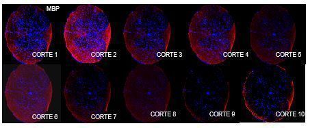
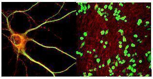
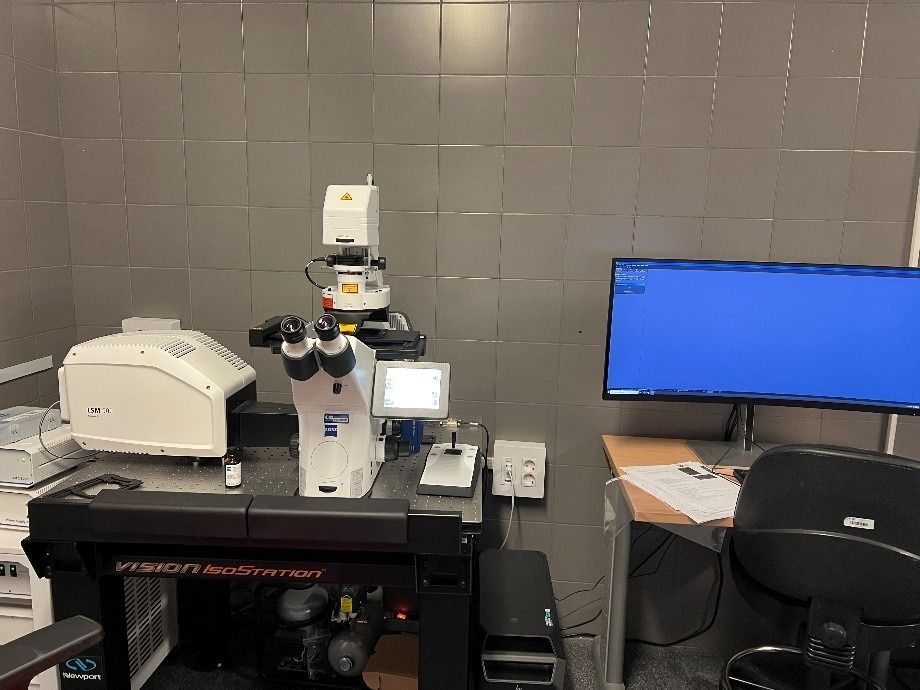
The IDIPHISA Advanced Optical and Confocal Microscopy Unit is equipped with:
LSM90 Confocal Microscope with Airyscan 2.0 (super-resolution module)
With a motorised stage, 4 solid-state laser lines directly connected to the scanning unit to avoid alignment issues, with the following power outputs:
- 405 nm laser (15 mW) 488 nm laser (25 mW)
- 561 nm laser (25 mW)
- 640 nm laser (15 mW)
4 detectors:
- 2 MA-PMT detectors for fluorescence with 16-bit Airyscan dynamic range
- 2 high-sensitivity units.
Increased resolution and SNR; 1 ESID detector for transmitted light.
Microscopio Confocal Leica SP5
with three laser lines, is optimised for image acquisition from samples, both fixed and in vivo, that do not require specific environmental conditions. This system has 3 excitation laser lines:
- Argon laser, exciting at the following wavelengths: 458, 477, 488, 496 and 514 nm.
- Helium/Neon laser (He/Ne), exciting at 543 nm.
- Helium/Neon laser (He/Ne) exciting at 633 nm.
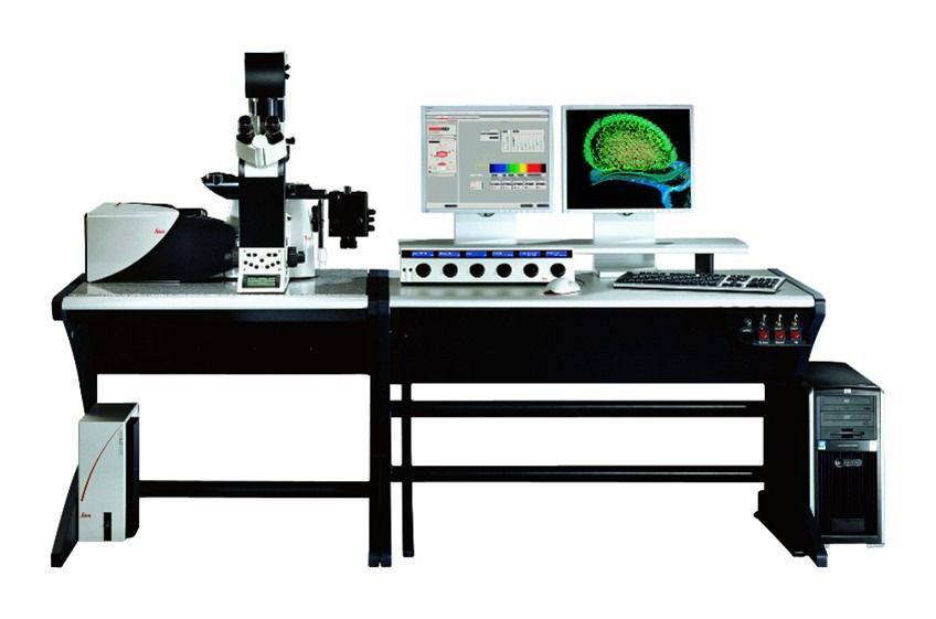
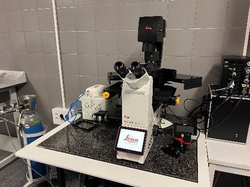
Microscopio Wide Field Thunder LEICA
- Microscopio Leica 5 LEDS
- Inverted microscope with 5x, 20x, 40x, 63x magnifications, with phase contrast and bright field.
- Motorised stage, CO2 incubator, temperature control, sCMOS camera (Leica K5)
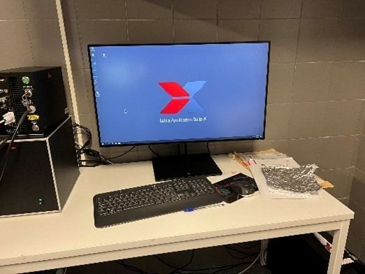
The unit also has:
- a citospin de Shandon to transfer cells grown in suspension onto slides, and the Unit also has 2 transmitted-light microscopes.
- a Leica transmitted-light microscope connected to a Leica MC190HD camera and a computer, enabling the acquisition of bright-field images such as Haematoxylin and other stains
Services Portfolio
The service offered by the Confocal Microscopy Unit is to assist the centre's research groups or visiting researchers in the process of obtaining images using Confocal Microscopy techniques, project planning, sample preparation, marker selection, image processing, quantification using specific software, data transfer, and image presentation and storage.
The Service maintains a fairly extensive stock of fluorochromes, which it acquires based on users' needs, and provides aliquots at low cost.
Services Portfolio
- Sample preparation.
- Sample processing for Fluorescence Microscopy
- Immunofluorescence.
- Sample visualisation and image capture on the Confocal Microscope.
- Data analysis, quantification, image processing, preparation of plates for publications.
- Advisory services.
FEES

The Unit's manager will control and operate the microscope and software. At no time may the user turn on or operate the Confocal Microscope, as its complexity requires qualified personnel specifically trained in its use.
Image transfers will be carried out by recording them onto a storage medium such as compact discs.
STAFF
- Scientific Coordinator: Dra. Margarita Sánchez-Beato
- Scientific Head of the Unit: Dra. Maria José Coronado
Tel: 91 191 67 56 - E-mail: confocal@idiphim.org
- Location: 3rd floor of the Research Area, Research Institute, Hospital Puerta de Hierro Majadahonda
SERVICE BOOKING FOR PUERTA DE HIERRO RESEARCHERS:
To check rates and use the service, you must make a prior booking at the Unit website (an internal user password is required).
SERVICE BOOKING FOR EXTERNAL USERS:
You may request the service by contacting the Head of the Unit by email in advance.